Osteochondral Defect
Background

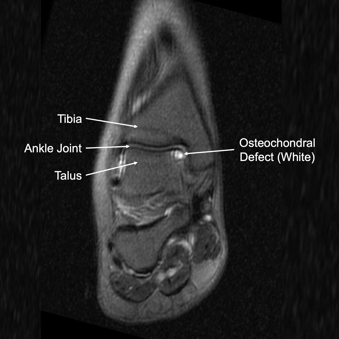
Ankle sprains are common, and we generally recover from them pretty well. However, sometimes damage is done to the osteochondral layer deep in side the joint. This layer consists of cartilage and subchondral bone (bone just below the cartilage). When this occurs, the damage can remain silent for months or years. Eventually, the cartilage begins to detach from the underlying unhealthy bone. The cartilage can then catch on adjacent surfaces and generate loose fragments that float around in the ankle joint. This causes inflammation, pain, swelling, difficulty with walking, and limitations in sports. These symptoms can be particularly troubling because affected patients are generally young, healthy, and active.
Nonsurgical Treatment
Nonsurgical treatment is largely in the form of symptom mitigation, meaning we are helping you to live with your symptoms, but not definitively curing the problem.
Helpful nonsurgical treatments include boot or brace immobilization, physical therapy for ankle strength and proprioception, non-steroidal anti-inflammatories, rest, ice, and activity modification.
If these fail to provide sufficient relief, Dr. Bohl will likely recommend an MRI, which is the only way to definitively diagnose an osteochondral defect.
Surgical Treatment
Dr. Bohl performs surgery for osteochondral defects through two minimally invasive incisions <0.5cm using an arthroscope.
The good news is that we have excellent, minimally invasive surgical treatments available for osteochondral defects.
If you have a diagnosed osteochondral defect and have failed conservative treatment, Dr. Bohl is likely to offer you an ankle scope. In this procedure, two tiny (<0.5cm) portals are made on the front of the ankle. Through one portal, a camera is inserted. Through the other portal, a shaver is inserted. These two instruments are used to visualize and remove the damaged cartilage and underlying subchondral bone. Tiny holes are then made in the remaining bone to release your body’s natural stem cells and activate them to form new cartilage.
After the procedure, you will be placed in a splint for two weeks, after which you can transition to a regular shoe and begin physical therapy.
Recurrent Osteochondral Defects and Revision Procedures
There are cases in which removal of the unhealthy cartilage and bone results in only a year or two of relief. In these cases, the body has failed to effectively form new healthy cartilage and patients experience persistent pain and swelling. A repeat MRI is needed, which generally shows additional unhealthy bone and cartilage. When this occurs, Dr. Bohl may recommend one of several options based on the amount of damage:
Moderate defects: Revision scope and subsequent reconstruction through a 3cm incision with bone graft and stem cells from a donor patient
Major defects: Bulk transplant of the osteochondral layer from a donor patient